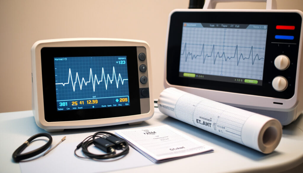
What Is an EKG? Test, Procedure, and Results Explained
Few medical tests cause as much quiet dread as an EKG — the word itself sounds clinical and cold. But the reality is almost anticlimactic: you lie still for a few minutes while 10 sticky electrodes listen to your heart’s electrical conversation, and what that conversation reveals is worth understanding before you ever need the test.
Duration: 5–10 minutes ·
Electrodes: 10 ·
Pain level: None ·
Typical cost: $50–$200
Quick snapshot
- Noninvasive test that records the heart’s electrical activity (NHS (UK public health authority))
- Also called an ECG (electrocardiogram) (NHS (UK public health authority))
- Typically takes 5–10 minutes from start to finish (Mayo Clinic (leading U.S. medical center))
- An abnormal EKG does not always mean heart disease — follow-up tests are often needed (American Heart Association (cardiology authority))
- Standard EKGs can miss arrhythmias that come and go, such as paroxysmal atrial fibrillation (American Heart Association (cardiology authority))
- Ordered when a patient reports chest pain, palpitations, or shortness of breath (Mayo Clinic (U.S. academic medical center))
- Routinely used for pre-surgical clearance and athlete screening
- Results are reviewed by a cardiologist who may recommend an echocardiogram, stress test, or Holter monitor (American Heart Association (heart health authority))
- Some clinics provide a preliminary reading before you leave
Five key specifications, one takeaway: an EKG is a short, low-cost snapshot of your heart’s electrical health — not a full diagnostic workup, but the most common starting point.
| Specification | Value |
|---|---|
| Full name | Electrocardiogram |
| Duration | 5–10 minutes |
| Pain | None |
| Cost | $50–$200 |
| Electrodes | 10 |
What is an EKG and what does it detect?
What does an EKG measure?
- An EKG records the electrical signals that make your heart beat — the same signals that coordinate each contraction of the upper and lower chambers (American Heart Association (cardiology authority)).
- It detects heart rhythm problems (arrhythmias), signs of blocked or narrowed arteries, and evidence of a past or current heart attack (NHS (UK public health service)).
- The test delivers no radiation and involves no needles or dyes (Cleveland Clinic (U.S. academic medical center)).
The implication: an EKG is not a full heart anatomy scan — it’s a functional recording of electrical activity. Structural problems like valve leaks or chamber size often require other imaging.
How does the test work?
- Ten electrodes in a standard 12-lead configuration capture the heart’s electrical impulse from 12 different angles (Cleveland Clinic (cardiology diagnostics center)).
- Six electrodes sit on the chest; four are placed on the limbs — one on each arm and one on each leg (NHS (UK public health authority)).
- The machine amplifies the tiny electrical signals and prints them as a waveform tracing on graph paper (StatPearls / NCBI (medical education reference)).
The trade-off: the machine captures only about 10 seconds of heart activity. That’s enough to detect persistent problems but can miss intermittent issues.
The 12-lead view is what makes an EKG so valuable — each angle gives a different “window” into the heart. A patient with chest pain might show ST-segment elevation in leads V2–V4, pointing directly to a blockage in the left anterior descending artery. The pattern saves lives precisely because it’s specific.
What this means: the specificity of the 12-lead view is what gives the EKG its diagnostic power in emergency settings.
What are three reasons a person would get an EKG?
To check heart rhythm irregularity
- Palpitations, a fluttering sensation in the chest, or a feeling that the heart is skipping beats are among the most common reasons physicians order an EKG (Mayo Clinic (cardiology department)).
- Atrial fibrillation, the most common arrhythmia, can be detected on a resting EKG if it’s present during the recording (American Heart Association (heart rhythm experts)).
To diagnose a heart attack
- Chest pain accompanied by shortness of breath, nausea, or jaw pain triggers an urgent EKG to check for a heart attack in progress (NHS (emergency cardiology guidance)).
- Certain waveform patterns — like ST-elevation — tell doctors which part of the heart muscle is affected and how urgently blood flow needs to be restored.
To evaluate symptoms like chest pain or shortness of breath
- Patients with unexplained shortness of breath, dizziness, or fainting spells often receive an EKG as a first-line test (UPMC (Pittsburgh academic health system)).
- Routine pre-operative evaluations and sports physicals also include an EKG for individuals with a family history of heart disease (Cleveland Clinic (preventive cardiology)).
The pattern: across all three reasons — rhythm, attack, symptom — the EKG serves as the fastest, cheapest first filter. It doesn’t confirm everything, but it tells the clinician where to look next.
A patient who walks into an ER with chest pain can have a 12-lead EKG recorded, transmitted to a cardiologist, and interpreted in under 5 minutes. That speed — not depth — is the EKG’s superpower.
The catch: the EKG’s speed makes it invaluable in emergencies, but its limitations mean it’s rarely the final word.
What should you not do before an EKG?
Avoid caffeine and smoking before test
- Caffeine — from coffee, tea, energy drinks, or chocolate — can increase heart rate and alter the natural rhythm, potentially producing a false reading (NHS (patient preparation guidance)).
- Smoking before the test also raises heart rate and blood pressure, which can mask baseline electrical activity (American Heart Association (pre-test guidance)).
Do not apply lotions or creams to chest
- Oily or greasy lotions, body oils, and talcum powder create a barrier between the skin and the electrode, interfering with signal quality (Cleveland Clinic (preparation instructions)).
- The NHS specifically advises avoiding body lotions, oils, or talc before an ECG (NHS (pre-test care)).
- In some cases, hair on the chest may be shaved in small patches to improve electrode adhesion (Mayo Clinic (clinical preparation)).
The catch: preparation is mostly about removing barriers — chemical barriers (lotions), physiological barriers (caffeine), and physical barriers (clothing). No fasting, no needles, no sedation.
What is an EKG and how long does it take?
What happens during the test?
- You remove clothing from the top half of your body and lie down on an examination table (NHS (procedure description)).
- A technician attaches 10 adhesive electrodes to your chest, arms, and legs — no pain, just a brief cool sensation from the gel (Cleveland Clinic (step-by-step guide)).
- You lie still, breathe normally, and do not talk for the duration of the recording to avoid muscle movement artifacts (Mayo Clinic (patient instructions)).
How long does the recording last?
- The actual electrical recording captures a standard 10-second strip of your heart rhythm (StatPearls / NCBI (clinical standards)).
- The total appointment — including setup, electrode placement, and cleanup — takes 5–10 minutes from start to finish (Mayo Clinic (duration estimate)).
- No recovery time is needed — you can return to normal activities immediately after the test (American Heart Association (post-test guidance)).
What this means: the test is shorter than most people expect. The setup takes longer than the recording itself, and the whole visit rarely exceeds 15 minutes including paperwork.
Is getting an EKG serious?
Are there any risks?
- The EKG is considered one of the safest medical tests in existence — it involves no radiation, no needles, and no contrast dyes (American Heart Association (safety statement)).
- Some people experience mild skin irritation or redness from the adhesive on the electrodes, which resolves within hours to a day (NHS (side effects)).
- Removing the electrodes may cause minor discomfort — similar to removing a bandage — but no pain during the test itself (American Heart Association (patient comfort)).
What does a serious EKG result mean?
- An abnormal EKG does not automatically mean heart disease — many non-cardiac factors (electrolyte imbalances, anxiety, even cold room temperature causing shivering) can produce abnormal-looking tracings (StatPearls / NCBI (artifact and error sources)).
- Abnormal results typically lead to follow-up tests such as an echocardiogram, stress test, or Holter monitor before any diagnosis is made (Mayo Clinic (diagnostic pathway)).
- A “normal” EKG is also not a guarantee of perfect heart health — it only shows the heart’s electrical activity during those 10 seconds (Cleveland Clinic (test limitations)).
The trade-off: an EKG carries almost zero physical risk, but it carries interpretive risk — false positives can cause anxiety, and false negatives can provide false reassurance. That’s why results are always read in context by a trained cardiologist.
An athlete with a thick heart muscle from years of training can show an “abnormal” EKG by population standards but be perfectly healthy. Without the clinical picture, the tracing alone is just a pattern — not a verdict.
The implication: context is everything — an EKG result must always be interpreted alongside symptoms, history, and additional testing.
How to prepare for an EKG: a step-by-step checklist
- Check with your doctor about medications. Tell your care team about all prescription drugs, over-the-counter medications, and supplements you take. Some medications can affect heart rate and rhythm (UPMC (medication guidance)).
- Avoid caffeine and nicotine for at least 2 hours before the test. Both are stimulants that can artificially raise your heart rate (NHS (preparation advice)).
- Skip lotions, oils, and talcum powder on your chest and limbs. These products reduce the electrode’s ability to pick up clear signals (Cleveland Clinic (pre-test skin care)).
- Wear a two-piece outfit. A shirt that opens in the front or a loose top makes it easy to expose your chest while keeping the rest of you covered (NHS (clothing advice)).
- Remove jewelry and metallic objects from your neck, wrists, and chest area before the recording begins (StatPearls / NCBI (pre-procedure checklist)).
- Stay still, breathe normally, and do not talk during the recording. Even slight movement can create electrical noise that obscures the real signal (Mayo Clinic (recording instructions)).
- Tell the technician if you feel cold. Shivering produces muscle artifacts that can distort the tracing. A blanket or warmer room temperature helps (StatPearls / NCBI (environmental factors)).
Why this matters: each step removes a source of noise. A clean recording means the cardiologist can read the true electrical pattern instead of guessing which blip came from a lotion barrier or a shaky arm.
Confirmed facts vs. what remains unclear
Confirmed facts
- An EKG is a noninvasive test that records the electrical activity of the heart (NHS).
- The test takes 5–10 minutes and is painless (Mayo Clinic).
- It carries no known medical risks and uses no radiation (American Heart Association).
- A standard 12-lead EKG uses 10 electrodes placed on the chest, arms, and legs (Cleveland Clinic).
What’s unclear
- An abnormal EKG does not always indicate heart disease; follow-up testing is typically required to confirm (American Heart Association).
- Intermittent arrhythmias — those that come and go — may not appear during the 10-second recording window (Cleveland Clinic).
- Certain conditions, such as electrolyte imbalances or anxiety, can mimic abnormal heart patterns on the tracing (StatPearls / NCBI).
- A “normal” EKG result does not guarantee perfect heart health — it only reflects the heart’s electrical activity during the brief recording period.
“An ECG is a quick and painless test that records the electrical activity of the heart. It is used to check the heart’s rhythm and find out if it is working normally.”
“During a resting ECG, you are usually asked to lie still and relax so that movement does not interfere with the recording. You can breathe normally but should not talk.”
— Mayo Clinic (cardiology patient education)
“The standard resting ECG procedure uses sticky patches or electrodes placed on the chest, arms, and legs. A typical resting ECG uses 12 electrodes: six on the chest and six on the limbs.”
— Cleveland Clinic (cardiology diagnostics center)
The pattern across all three sources is consistent: the EKG is simple, fast, and safe — but its interpretation depends on a skilled clinician who knows what to look for and what the test cannot see. For the patient, understanding the limits of an EKG is just as important as understanding what it can detect.
mdmedicalclinics.com, northwestfamilyclinics.com, heart.org, ahajournals.org, capitolcardiology.com, youtube.com
For a more in-depth look at how the test is performed, you can refer to this detailed guide on EKG procedure.
Frequently asked questions
What is the difference between an EKG and an ECG?
None. EKG and ECG are abbreviations for the same test — electrocardiogram. “EKG” comes from the German spelling (Elektrokardiogramm) and is commonly used in the United States, while “ECG” is more common in the UK and elsewhere (American Heart Association).
Can an EKG detect a heart attack?
Yes, an EKG can detect a heart attack in progress or signs of a past heart attack. Specific waveform patterns — such as ST-segment elevation — indicate that a portion of the heart muscle is not receiving enough oxygen (NHS).
How often should I have an EKG?
There is no standard frequency for everyone. Healthy adults without symptoms or risk factors may not need a routine EKG. Your doctor may recommend one every 1–3 years if you have high blood pressure, diabetes, a family history of heart disease, or other risk factors (Cleveland Clinic).
Do I need to fast before an EKG?
No. Fasting is not required for a standard resting EKG. However, you should avoid caffeine and smoking for at least 2 hours before the test, as both can affect heart rate and rhythm (UPMC).
Can stress affect EKG results?
Yes. Acute stress and anxiety can raise heart rate and blood pressure, which may produce changes in the EKG tracing. Some people also hyperventilate when anxious, which alters electrolyte levels and can mimic abnormal patterns (StatPearls / NCBI).
What do the electrodes do?
Each electrode acts as a tiny sensor that detects the electrical impulses generated by your heart muscle as it contracts. The 12 electrodes together provide a 360-degree view of the heart’s electrical activity, allowing the machine to map the direction and timing of each impulse (Cleveland Clinic).
Is an EKG covered by insurance?
Most health insurance plans in the United States cover an EKG when it is ordered by a physician for a medical reason — such as chest pain, palpitations, or pre-surgical screening. Out-of-pocket costs typically range from $50 to $200 without insurance (UPMC).
For the patient facing an EKG for the first time, the choice is clearer than it feels: a few minutes of lying still for a recording that could catch a serious problem before it becomes an emergency — or miss nothing at all if your heart happens to be on its best behavior. That’s the honest trade-off of a 10-second snapshot, and it’s exactly why your doctor pairs it with your symptoms, your history, and often a second test before drawing conclusions.
If you’re curious about how other common medical tests compare, check out our explainer on What Is Strep Throat? or our guide to What Is Body Dysmorphia? — both follow the same evidence-first approach.
More related posts
 How to Deal with a Narcissist: Expert Strategies and Tips
 What Does IMO Mean in Texting? Definition and Usage
 How to Stop Tooth Pain Fast: 5 Immediate Home Remedies
 Cast of Hysteria TV Show: Who’s Who in Peacock Horror Comedy
 Big Little Lies Cast: Complete Seasons 1, 2 & 3 List
 Alabama Football Schedule 2025: Dates, Odds, Playoff Path
 Tank Tops for Women: Best Styles & Top Picks 2024